Systemic lupus erythematosus is a chronic inflammatory autoimmune disease with a broad spectrum of symptoms, whose origin is multifactorial. The disease presents with a diverse clinical morphology and different organic manifestations, ranging from localized cutaneous lupus erythematosus to the severe systemic form.
In order to make the diagnosis of patients in a standardized way, criteria for classification of the disease have been defined. On the one hand, there are classifications from the rheumatologic point of view, which are based on the symptomatic manifestations and allow the assessment of systemic involvement.
Similarly, a classification criterion has been established from the dermatological point of view, which categorizes the disease according to the morphology of the cutaneous manifestations and according to the acuity. In this criterion we have, cutaneous subacute lupus erythematosus, acute cutaneous and chronic cutaneous. The latter includes chronic discoid variants, lupus profound and lupus tumidus. Then, depending on the skin compartment involved, histologic classification may be epidermal, epidermal, dermal, or subcutaneous lupus erythematosus.
Points of interest for the diagnosis of lupus
This disorder may be difficult to determine, and there is no specific test to diagnose it. For this, different laboratory tests are used to detect changes or conditions in the body, which can be caused by lupus. Each test provides information about the disease.
Recommended laboratory tests are C-reactive protein, complement level, urine, antinuclear antibodies, anti-DNA antibodies, anti-SSA/Ro antibodies, anti-Smith antibodies, anti-phospholipid antibodies. Skin and kidney biopsies are also recommended.
Histology is an important component in confirming the diagnosis of this disorder. However, histopathologic changes cannot be assigned to a specific diagnosis because inflammatory patterns are similar in other skin manifestations. For this reason, conclusions should not be drawn solely on the basis of histological findings. Correlation of clinical and histologic findings is essential.
Preparation of tissue sample for histopathological examination
To confirm the presence of cutaneous lupus and rule out another skin condition that can cause similar symptoms, a skin biopsy is recommended by the medical professional. If doctors suspect kidney involvement because of symptoms or detect an abnormal result after a urine test, they also recommend a kidney biopsy.
Histologic analysis can establish the level of inflammation and any other tissue damage. To perform this study, the tissue sample must be carefully obtained and manipulated, then fixed, usually in formalin; then the sample must be dehydrated by means of increasing ethanol concentrations and then rinsed with xylene. Subsequently, the tissue is infiltrated with paraffin, and then included in a paraffin block, which will allow the sample to be cut into a microtome.
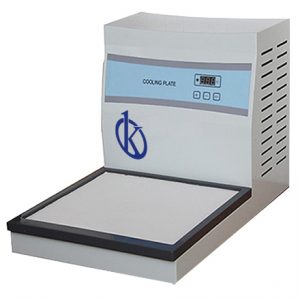
Once the sample has been included in the paraffin block, cooling of the paraffin block is essential, to obtain sections of good quality tissue. This is done with equipment called cooling plate, which is an essential device in histology laboratories. An adequate cooling process of the paraffin block will facilitate the obtaining of thinner tissue sections and avoid the appearance of cracks. The latter may lead to total sample loss.
Kalstein cooling plates
In Kalstein we have for sale cooling plates of the models YR440 and YR446. These are two models with different dimensions, which allow you to adjust to the different needs of the laboratory. Kalstein equipment has a cryoplate adapted to an inverter type compressor, which facilitates rapid cooling. In addition, they are designed with a Teflon coating, which allows easy cleaning. They also offer the possibility to adjust the temperature, by digital regulation and allow the simultaneous cooling of approximately 96 paraffin blocks. For more information on Kalstein cooling plates, visit HERE