Pathological anatomy is the science that studies the pathophysiological and morphological alterations of the disease. It studies the disease at an organic, tissue, cellular, subcellular, and molecular level. It is divided into:
- General Pathological Anatomy: Study principles common to groups of diseases, which allow the development of a doctrine of more or less universal validity.
- Special Pathological Anatomy or Surgical Pathology: Study the pathophysiological and morphological bases of each particular disease.
What types of tests are performed in pathology?
The main objective of pathological anatomy is not only to study the evolution and causes of a disease, but also to make a prognosis of the effects that different diseases can have on human beings. To do this, tumor cells and body tissues are analyzed.
Thus, within pathological and cytodiagnostic studies, two types of tests are differentiated: pathological and diagnostic. In the first of them, a sample of the tissue of any organ is analyzed with the intention of identifying the type of cell and what treatments would be useful to combat the alteration. As for diagnostic tests, they are also known as biopsy and their main objective is to obtain a sample of organic fluid or tissue to study the existence of a disease, with the intention of determining a specific diagnosis.
What are the main procedures performed in pathological anatomy?
Pathological anatomy performs various methods depending on the type of tissue and the type of diagnosis that is presumed, among them we can mention:
- Histopathology: It is the microscopic examination of a sample using histological techniques. Stains provide a specific diagnosis based on morphology and is the core skill of histology.
- Immunohistochemistry: It is based on the use of antibodies to detect the presence, abundance and localization of specific proteins. This technique is critical for distinguishing between disorders with similar morphology and also for characterizing the molecular properties of some cancers.
- In situ hybridization: Specific DNA and RNA molecules can be identified in sections using this technique.
- Cytopathology: It is the examination of free cells spread in a petri dish using cytology techniques
- Electron microscopy: It consists of the examination of tissue with an electron microscope that allows greater magnification, allowing the visualization of organelles within the cell.
- Tissue cytogenesis: It is based on the visualization of chromosomes to identify genetic defects such as chromosome translocation.
- Immunophenotyping: It consists of determining the immunophenotype of cells using cytometric techniques. (measure cells). It is useful for diagnosing different types of leukemia and lymphomas.
Main equipment used in pathological anatomy
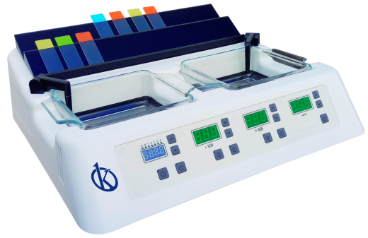
Among the equipment most used in this science, we have: microtomes, microscopes, tissue processor, water baths for tissues, cryostats, cooling plates, trimmer and paraffin dispenser, slide dryers, tissue inclusion systems, systems of automatic staining for slides, among others.
At Kalstein we are MANUFACTURERS, so you can BUY everything you need for your pathology laboratory at excellent PRICES. That is why we invite you to take a look at the Products menu. HERE